X-rays and interaction with crystalline matter
X-rays are electromagnetic radiation with wavelength λ ≈ 0.5–2.5 Å (50–250 pm), comparable to interatomic distances in crystals (C–C bond ≈ 1.54 Å, O–H bond ≈ 0.96 Å). This commensurability between λ and unit-cell parameters makes X-ray diffraction possible.
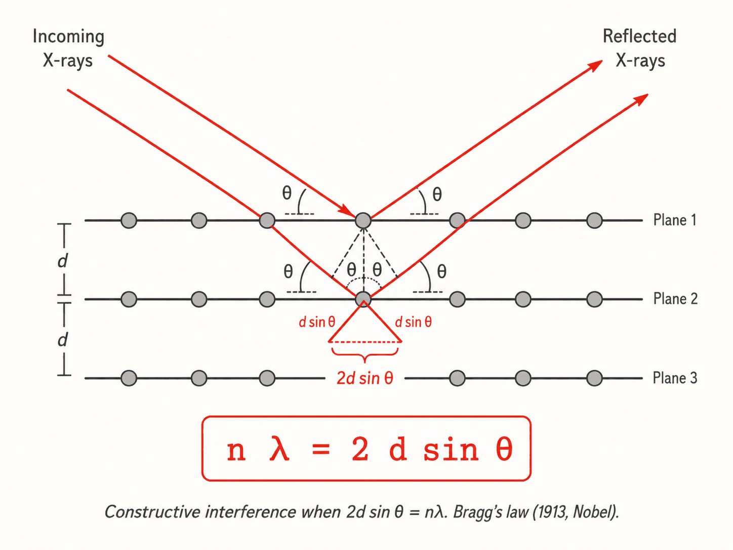
When an X-ray beam hits a crystal, each atom scatters radiation coherently (Thomson scattering). The scattered waves from different lattice planes interfere constructively when the optical path difference is an integer multiple of λ — this is Bragg's condition.
Bragg's law
William Henry Bragg and William Lawrence Bragg established in 1913 the fundamental relation:
n λ = 2 d sin θ
where: - n is the diffraction order (positive integer, most often n = 1), - λ is the X-ray wavelength (Å), - d is the interplanar spacing (distance between two parallel (hkl) planes, in Å), - θ is the half-angle of diffraction (Bragg angle, in °).
Bragg's law allows measuring d from known λ and observed θ, or determining λ from known d (monochromator). It is valid for wavelengths satisfying λ ≤ 2d (otherwise sin θ > 1, impossible).
For laboratory X-rays (Cu tube, K_α, λ = 1.5406 Å), Bragg's law is directly applicable to the majority of interplanar spacings (1–10 Å) in inorganic and organic solids.
Single crystals vs. powders
Single-crystal X-ray diffraction (SC-XRD):
A single crystal (ideal size: 0.05–0.5 mm) is mounted on a goniometer. The crystal angle varies in 3D during acquisition (ω-scan method). Diffraction spots (hkl reflections) are recorded; their positions and intensities allow solving the complete crystal structure (atomic positions, thermal parameters, bonds). The reference method for structural elucidation of small organic molecules and inorganic compounds.
Powder X-ray diffraction (PXRD):
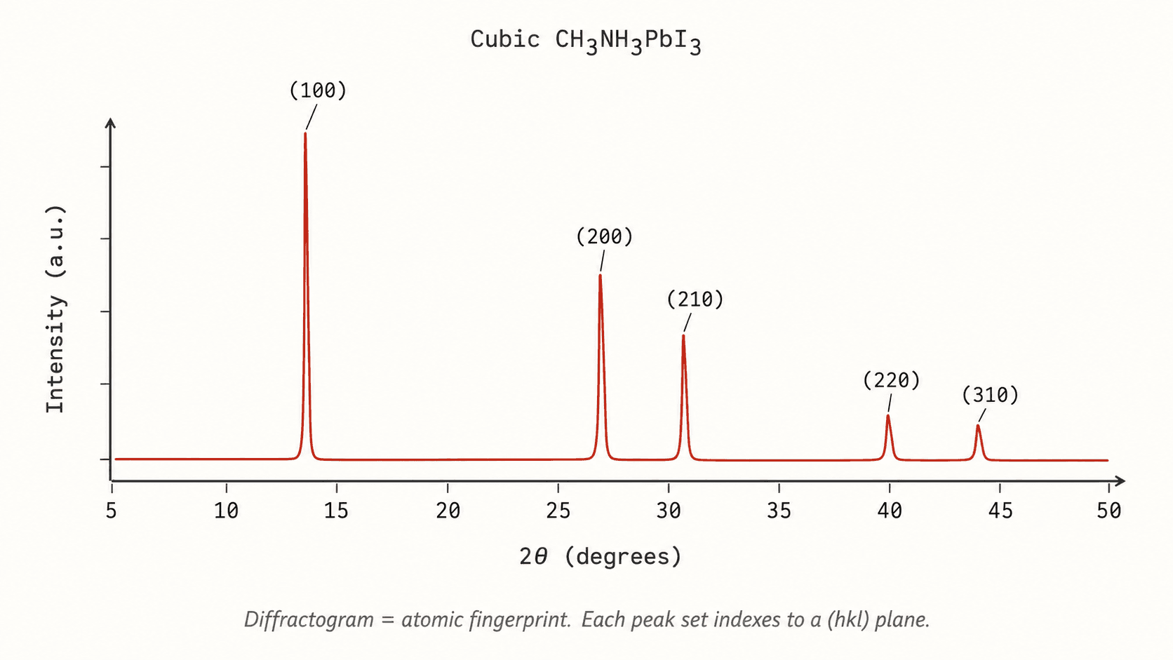
The sample is a polycrystalline powder. The random orientation of crystallites means all hkl reflections are simultaneously present. The detector records intensity as a function of 2θ (diffractogram): each family of planes gives a peak at its Bragg angle. Applications:
- Phase identification by comparison with databases (ICDD-PDF4+).
- Quantification of mixtures (Rietveld method).
- Crystallite size measurement (Scherrer formula: τ = K·λ / (β·cos θ), where β is the full width at half maximum).
| Method | Advantages | Limitations |
|---|---|---|
| SC-XRD | Complete 3D structure, sub-Å accuracy | Requires a quality single crystal |
| PXRD | Ordinary powder, fast identification | Less 3D structural information |
Structural applications: from DNA to perovskites
Discovery of the double helix (1953): X-ray fiber diffraction patterns from DNA obtained by Rosalind Franklin (Photo 51) provided key parameters (helix pitch 34 Å, diameter 20 Å) that Watson and Crick used to build the double-helix model. B-DNA has a 3.4 Å spacing between stacked bases, detectable at θ ≈ 13° for λ = 1.54 Å.
Perovskites ABX₃: The perovskite structure (e.g. BaTiO₃, CaTiO₃, methylammonium lead iodide MAPbI₃) is determined by PXRD. The unit-cell parameter a is measured from Bragg peak positions, and distortions (octahedral TiO₆ tilting) are quantified by Rietveld refinement. Halide perovskites are at the heart of third-generation solar cells with efficiencies > 26%.
Zeolites and MOFs: X-ray diffraction is indispensable for characterizing the cages and channels of porous materials used in catalysis and separation.
Proteins: SC-XRD (or high-resolution cryo-EM) provides atomic coordinates deposited in the Protein Data Bank (PDB, > 200 000 structures). Resolution is expressed in Å: at 2 Å side chains are visible; at 1 Å hydrogen positions become accessible.
Synchrotron and recent developments
Synchrotron sources (ESRF Grenoble, SOLEIL Paris) produce an X-ray photon flux 10¹²× higher than a laboratory tube, with tunable λ. They enable:
- Time-resolved diffraction (monitoring phase transitions in ms).
- Anomalous diffraction (MAD/SAD) to locate heavy metals in proteomics.
- Micro-beam (1 μm) to study tiny single crystals or individual grains.
Transmission electron diffraction (TED) and 3D electron diffraction (3D-ED / MicroED) allow structure solution from crystals of a few hundred nm, inaccessible to classical SC-XRD.
Safety and practical aspects
X-rays are ionizing radiation. Laboratory diffractometers are equipped with full shielding and interlocks (automatic cut-off on opening). Doses received in normal use (closed cabinet) are negligible. Synchrotrons require concrete shielding and dosimetry badges.
To prepare a PXRD sample: grind finely (< 10 μm), load into a sample holder (silicon flat or Lindemann capillary), minimize preferred orientation.