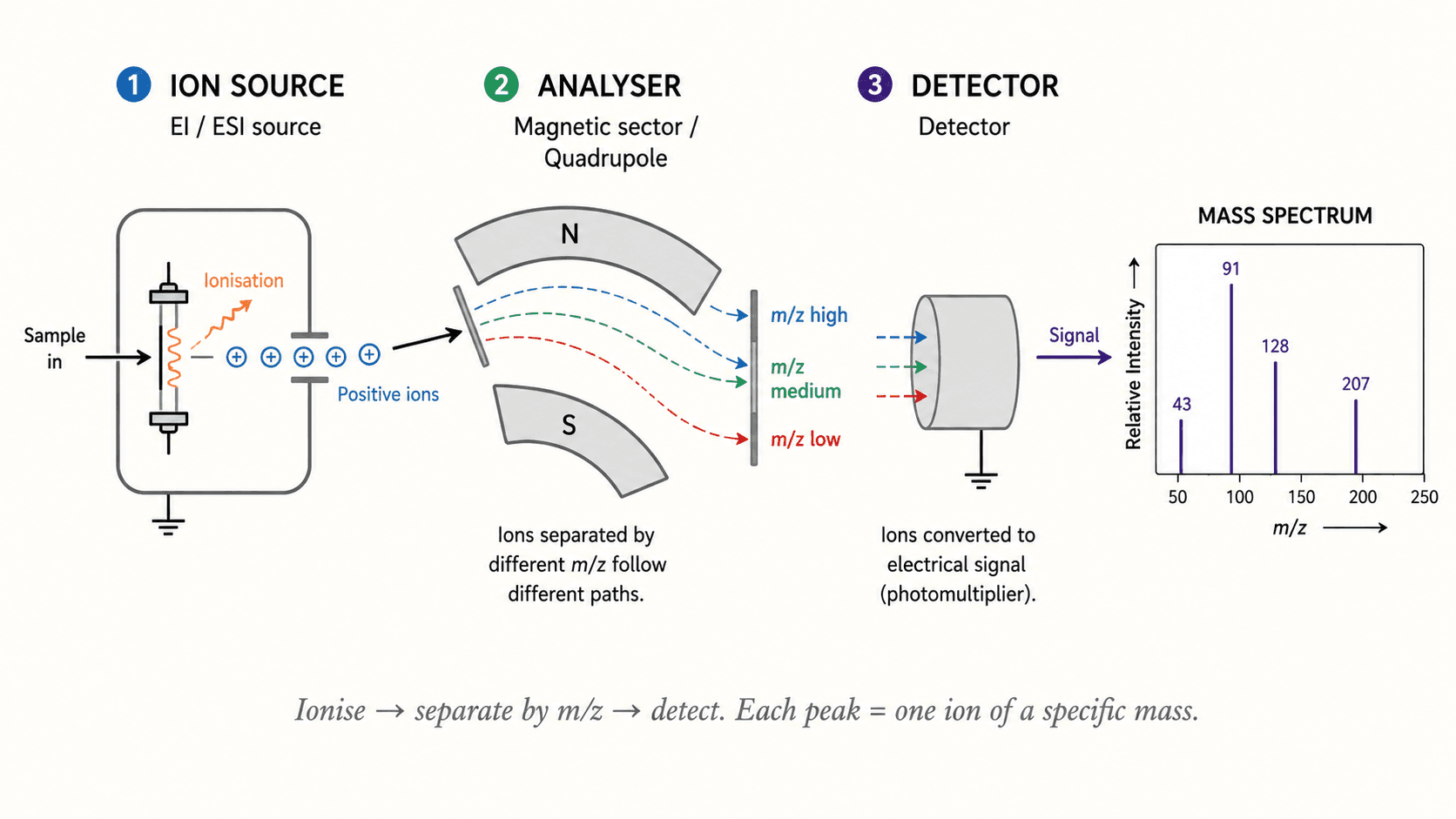
Principle and instrumentation
Mass spectrometry (MS) determines the mass-to-charge ratio (m/z) of gas-phase ions. The instrument has three parts:
1. Ionization source: produces ions from the sample. 2. Mass analyzer: separates ions by m/z. 3. Detector: measures the abundance of each ion.
A mass spectrum is a histogram of ions by m/z. Intensity is expressed as relative abundance: the base peak (100%) is the most abundant ion; all others are normalized to it.
Ionization modes
The choice of source determines the internal energy deposited in the molecule and thus the degree of fragmentation:
- Electron ionization (EI, 70 eV): energetic electron bombardment → radical cation M⁺·; abundant characteristic fragmentations. Suitable for volatile, thermally stable molecules.
- Chemical ionization (CI): reagent gas (CH₄, NH₃) → [M+H]⁺ or [M−H]⁻; soft ionization. Useful for molecular mass when EI gives a weak M⁺·.
- Electrospray (ESI): solution → spray → desolvation → [M+nH]ⁿ⁺. Soft, ideal for proteins and polymers. Gives multiply charged ions.
- MALDI (matrix-assisted laser desorption/ionization): co-crystallization with a UV-absorbing matrix → [M+H]⁺ ions of high mass. Standard in proteomics.
The molecular ion and exact mass
In EI, the molecular ion M⁺· corresponds to the molecule having lost one electron. Its m/z gives the nominal mass (sum of most abundant isotopic masses). Using high resolution (HR-MS, FWHM < 5 ppm), the exact monoisotopic mass is measured, enabling molecular formula determination.
Example: the exact mass of C₆H₆ (benzene) is 78.0469 Da. If HR-MS gives m/z = 94.0418, the formula C₆H₅F (fluorobenzene, theoretical mass 94.0425) is consistent.
The nitrogen rule: a molecule with an odd number of N has an odd nominal molecular mass. Useful to quickly identify amines.
The isotope pattern of the molecular ion reveals Cl or Br: - Cl: M and M+2 in 3:1 ratio (³⁵Cl : ³⁷Cl = 75.8 : 24.2%). - Br: M and M+2 in ~1:1 ratio (⁷⁹Br : ⁸¹Br = 50.7 : 49.3%).
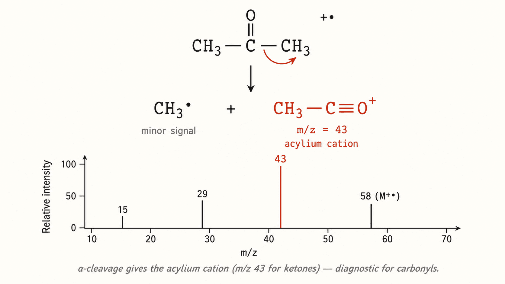
Characteristic fragmentations
Fragmentation occurs by well-defined mechanisms. The key ones to know are:
| Fragmentation | Loss (Δm) | Diagnostic |
|---|---|---|
| α-cleavage | variable | Ketones, aldehydes (acylium R–C≡O⁺) |
| McLafferty rearrangement | 58 (for Ph-CO–CH₂CH₂CH₃) | γ-H + carbonyl |
| Loss of H₂O | 18 | Alcohols, acids |
| Loss of CO | 28 | Aldehydes, phenols |
| Loss of CH₃ | 15 | Terminal methyl |
| Loss of Cl | 35 | Chlorides |
α-cleavage cuts the bond α to the heteroatom or charge: ketones R–CO–R' give acyliums R–C≡O⁺ (m/z = M − R') and R'–C≡O⁺ (m/z = M − R).
The ion at m/z = 77 is characteristic of the phenyl cation C₆H₅⁺; m/z = 91 corresponds to the tropylium ion C₇H₇⁺ (very stable, 6π aromatic), a signature of benzylic compounds.
GC-MS coupling
Gas Chromatography – Mass Spectrometry (GC-MS) coupling separates a mixture by GC (capillary column, 30 m × 0.25 mm, polysiloxane stationary phase) then analyzes each fraction by MS (EI source). The total ion chromatogram (TIC) gives a profile vs. retention time; each peak yields a mass spectrum that can be compared to libraries (NIST 20, Wiley).
Typical conditions: injection temperature 250 °C, temperature program 50 °C → 280 °C at 10 °C/min, He flow 1 mL/min. GC-MS is the reference technique for environmental trace analysis, anti-doping control and perfumery.
For non-volatile or thermolabile molecules, LC-MS/MS (HPLC-ESI-triple quadrupole) is preferred: precursor ion selection (Q1), fragmentation (Q2, collision-induced dissociation), fragment detection (Q3) → SRM mode, highly selective.
Interpreting a spectrum — method
1. Locate M⁺· (or [M+H]⁺ in CI/ESI) and note its m/z. 2. Analyze the isotope pattern for Cl, Br, S. 3. Calculate the degree of unsaturation: DU = (2C + 2 + N − H − X) / 2. 4. Identify losses from M: −15 (CH₃), −18 (H₂O), −28 (CO or C₂H₄), −45 (OC₂H₅). 5. Note characteristic m/z ions (77, 91, 105…). 6. Propose a structure consistent with all observations and confirm by NMR.